RMC ATUMtome for Array Tomography
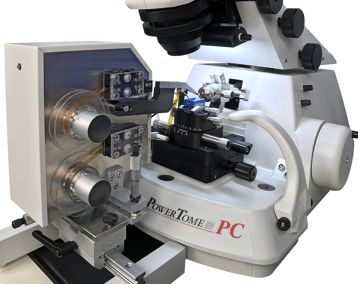
RMC ATUMtome is a unique ultramicrotome for fully automated collection of sections directly onto tape rolls and is a valuable tool for many other areas of research in biology, pathology and material sciences where three-dimensional image data of specimens is required. Originally developed at Harvard University in the lab of Jeff Lichtman for the collection of thousands of sections to reconstruct neuronal networks and pathways by array tomography.
The normally tedious process of collecting many serial sections is fully automated enabling ultra-thin sections with a thickness down to 30nm to be collected on 8mm wide Kapton tape for array tomography using SEM imaging and subsequent 3-D reconstruction.
Read about the advantages of ATUMtome in the Microscopy and Analysis article (Webster, P. et al 2015) M&A ATUMtome
- Fully automated high vacuum carbon coater
- PowerTome PTPCZ ultramicrotome with live video and computer control
- ATUM continuous tape feed mechanism with PC control software
- Air-activated anti-vibration table including
- ATUM attachment interface with x-y-z fine control positioning of tape/section pick-up head
- Silent air compressor for anti-vibration table
Environmental chamber for prevent turbulent air movement - Anti-static device for charge elimination on diamond knife edge
- Ergonomic laboratory chair
- 4mm DiATOME diamond knife, 35 degree for room temperature ultra-thin sectioning (order separately)
- Diamond knife boat water level control system
- Wafer workstation for cutting and organising tape strips with sections
- Start-up supply of Kapton tape
- Easy upgrade to add high resolution sputter coating
Working with ATUMtome
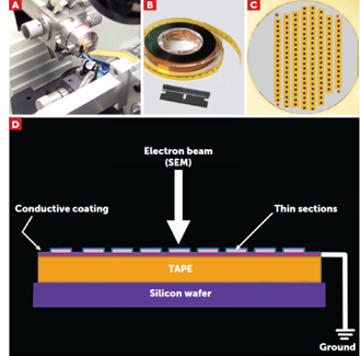
Section collection is carried out automatically using the ATUMtome (A). The tape with sections attached is taken from the spool and cut into small strips (B) and attached to a 4” silicon wafer (C). This shows how the strips of tape are mounted onto the silicon wafer, with metal TEM specimen grids positioned at the corners of the wafer. The grids act as markers for automated image collection (Hayworth, et al., 2014). The sections on the silicon wafer can be imaged using epifluorescent illumination (not shown), or with by SEM (D). Carbon coating lay below allows SE imaging, whilst for BSE imaging carbon is deposited over the sections.
Hayworth, K.J., Morgan, J.L., Schalek, R., Berger, D.R., Hildebrand, D.G. & Lichtman, J.W. (2014). ImagingATUM ultrathin section libraries with WaferMapper: a multi-scale approach to EM reconstruction of neural circuits. Front Neural Circuits 8, 68.
RMCATUM308-15. Uncoated Kapton tape for use on RMC ATUM, 8mm wide by 15 metres (each)
RMCATUM308-30. Uncoated Kapton tape for use on RMC ATUM, 8mm wide by 30 metres (each)
10-008140. Micro-Tec silicon wafer, Ø4 inch/100mm, 525µm thickness (each)
ATUM continuous tape feed mechanism with PC control software
PowerTome PCZ ultramicrotome
Air-activated anti-vibration table including ATUM attachment interface with x-y-z fine control positioning of tape/section pick-up head
Silent compressor for anti-vibration table
Environmental chamber
Anti-static device
Ergonomic lab chair
4mm diamond knife, 35 degree for room temperature ultra-thin sectioning, mounted in large-cavity blue anodized holder
Water level control system
Wafer workstation
Start-up supply of Kapton tape
Four 4” diameter silicon wafers
Many current users are working with the ATUMtome for neuroscience research, but this is far from the only application. Research
into biological systems and cell organelles as well as new materials research are being imaged using this array tomography technique. The ATUMtome’s unique ability to collect hundreds to thousands of sections on a continuous tape opens the door for
use in many serial section applications, some of which are yet to be explored.
The system can be used in correlative microscopy applications involving, for example, light and scanning electron microscopy to identify regions of interest and map nanoparticles inside organs and tumors. It can also benefit users who want to image whole cells and correlate the 3D distribution of specific proteins within these cells. Stored sections can be immunolabled multiple times for examination under epifluorescence illumination.
As research continues to transition from 2D to 3D imaging, there is a growing requirement to increase efficiency in imaging thousands of sample sections. By addressing this need, as well as the need to retain samples for future analysis, the ATUMtome is an exciting tool to consider. This is especially so among scientists who have wanted to carry out 3D reconstruction but were held back because of the impractical effort it would take to handle the many sections required.
Electrical input
110 – 240 VAC 50/60 Hz, output: 255 watts
Shipping Specifications
PowerTome crate: 865mm x 560mm x 660mm , 54.5kg
ATUM/Accessories: 660mm x 660mm x 660mm, 45.5kg
Computer: 660mm x 660mm x 305mm, 11.5kg
Workstation: 355mm x 355mm x 355mm , 9.0kg
Environmental chamber: 1550mm x 205mm x 205mm, 15.5kg
Slide Rail: 1220mm x 100mm x 100mm, 1.8kg
Compressor: 430mm x 380mm x 380mm , 19.5kg
Lab chair: 685mm x 685mm x 405mm, 18kg
Anti-vibration table: 1295mm x 1015mm x 940mm, 257.5kg
Total weight: 432kg
RMC Boeckeler is a specialist manufacturer of ultramicrotomes, microtomes and related instruments for the TEM and LM markets with a history dating back to 1941.
Based in Tucson Arizona in the USA, RMC Boeckeler is a privately owned. Their instruments are used globally in many fields including materials science and cell biology, with special emphasis in specimen preparation for 3D electron microscopy solutions.
Ordering information:
ATUMtome
Complete system (add Diatome knife – see below)
-
ATUMtome – Automated Tape Collecting Ultramicrotome
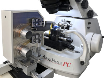
ATUMtome : developed at the Lichtman Lab at Harvard University the Automated Tape Collecting Ultramicrotome automates the section collecting process, improving work flow for imaging.
Price On Request £0.00 Add to basketATUMtome – Automated Tape Collecting Ultramicrotome
ATUMtome : developed at the Lichtman Lab at Harvard University the Automated Tape Collecting Ultramicrotome automates the section collecting process, improving work flow for imaging.
£0.00Price On Request
DiATOME diamond knife – new and resharpening
-
DiATOME ultra 45° diamond knife
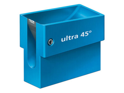
DiATOME ultra 45° diamond knife for ultra thin sectioning at ambient temperature
Price On Request £0.00 Select options This product has multiple variants. The options may be chosen on the product pageDiATOME ultra 45° diamond knife
DiATOME ultra 45° diamond knife for ultra thin sectioning at ambient temperature
£0.00Price On RequestSelect options This product has multiple variants. The options may be chosen on the product pageAdd to QuoteWithin your QuoteAdd to Quote -
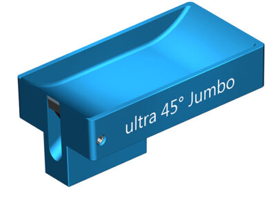
DiATOME ultra 45° Jumbo diamond knife
DiATOME ultra 45° Jumbo diamond knife for ultra thin sectioning at ambient temperature with large boat
Price On Request £0.00 Select options This product has multiple variants. The options may be chosen on the product pageDiATOME ultra 45° Jumbo diamond knife
DiATOME ultra 45° Jumbo diamond knife for ultra thin sectioning at ambient temperature with large boat
£0.00Price On RequestSelect options This product has multiple variants. The options may be chosen on the product pageAdd to QuoteWithin your QuoteAdd to Quote
ATUMtome accessories and consumables
-
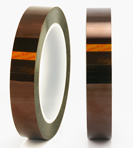
Uncoated Kapton tape, 33 metre
Uncoated Kapton tape – 33 metres
Price On Request £0.00 Add to basket -

Uncoated Kapton tape glow discharged, 33 metre
Uncoated Kapton tape – glow discharged – 33 metres
Price On Request £0.00 Add to basketUncoated Kapton tape glow discharged, 33 metre
Uncoated Kapton tape – glow discharged – 33 metres
£0.00Price On Request -
ATUMtome tape reel
ATUMtome Tape Reel
Price On Request £0.00 Add to basket -
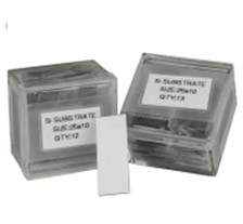
Silicon wafer substrates 10 x 25mm (pack of 13)
Silicon wafer substrates 10 x 25mm for standard knife boats – 1 pack of 13
Price On Request £0.00 Add to basketSilicon wafer substrates 10 x 25mm (pack of 13)
Silicon wafer substrates 10 x 25mm for standard knife boats – 1 pack of 13
£0.00Price On Request -

Double-sided carbon conductive tape, 25mm x 5m
Double Sided Carbon Conductive Tape- 25mm x 5m
Price On Request £0.00 Add to basketDouble-sided carbon conductive tape, 25mm x 5m
Double Sided Carbon Conductive Tape- 25mm x 5m
£0.00Price On Request -

Double-sided carbon conductive tape, 12mm x 5m
Double Sided Carbon Conductive Tape- 12mm x 5m
Price On Request £0.00 Add to basketDouble-sided carbon conductive tape, 12mm x 5m
Double Sided Carbon Conductive Tape- 12mm x 5m
£0.00Price On Request -

Carrier box 4 inch diameter wafers (capacity 25)
Carrier box for 25 numbers of 4 diameter wafers
Price On Request £0.00 Add to basketCarrier box 4 inch diameter wafers (capacity 25)
Carrier box for 25 numbers of 4 diameter wafers
£0.00Price On Request -

Carrier box for one 4 inch diameter wafer
Carrier box for 1 number of 4 diameter wafer
Price On Request £0.00 Add to basketCarrier box for one 4 inch diameter wafer
Carrier box for 1 number of 4 diameter wafer
£0.00Price On Request -

4 inch diameter silicon wafer (each)
4 Diameter Silicon Wafer – per piece ¹
Price On Request £0.00 Add to basket -
Large silicon wafer substrates 25 x 25mm (pack of 13)
Large Silicon Wafer substrates25 x 25mm- 1 pack of 13
Price On Request £0.00 Add to basketLarge silicon wafer substrates 25 x 25mm (pack of 13)
Large Silicon Wafer substrates25 x 25mm- 1 pack of 13
£0.00Price On Request -

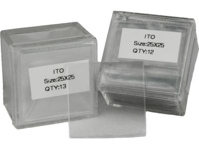
ITO (Indium Tin Oxide) coated glass substrates (pack of 13)
ITO (Indium Tin Oxide) coated glass substrates25 x 25 mm- 1 pack of 13
Price On Request £0.00 Add to basketITO (Indium Tin Oxide) coated glass substrates (pack of 13)
ITO (Indium Tin Oxide) coated glass substrates25 x 25 mm- 1 pack of 13
£0.00Price On Request
